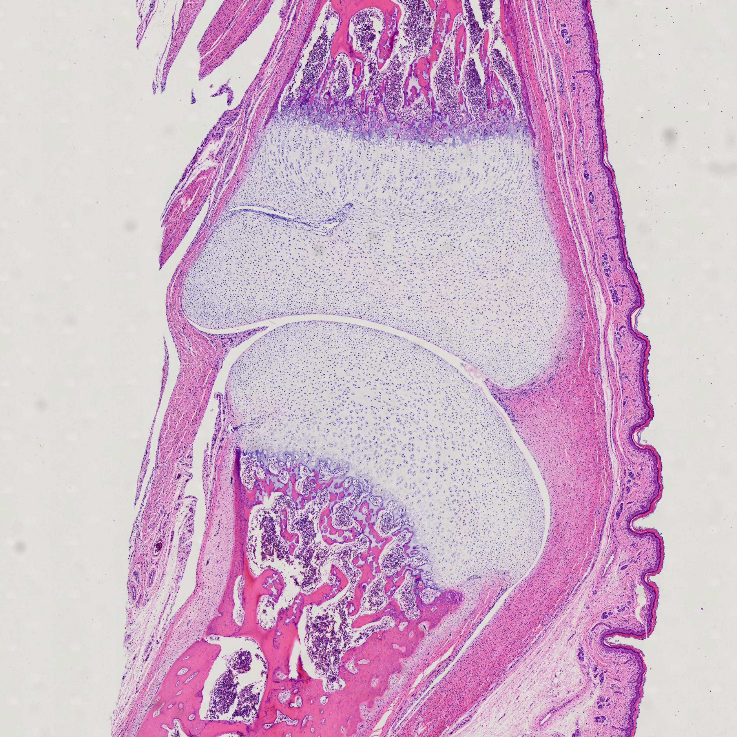
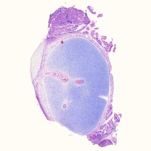
Endochondral Bone Development, human, H.E. Stain
The primary site of ossification can be seen in the diaphysis.
The epiphyseal plates continuously provide for the lengthening of the bone.
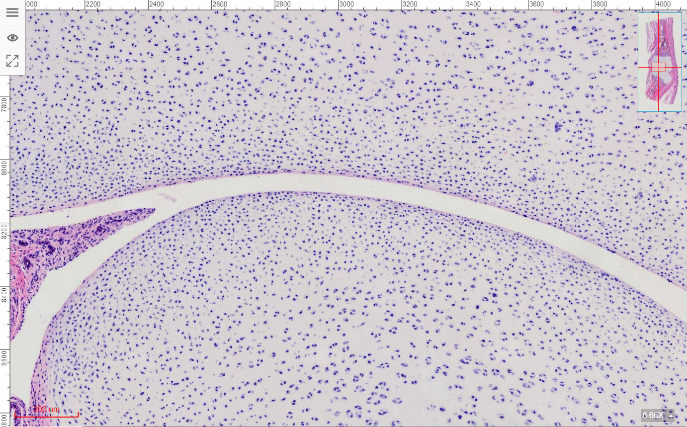
Resting Zone (Reserve) – quiescent chondrocytes
Proliferation Zone – rapidly dividing chondrocytes that form longitudinal columns of stacked cells
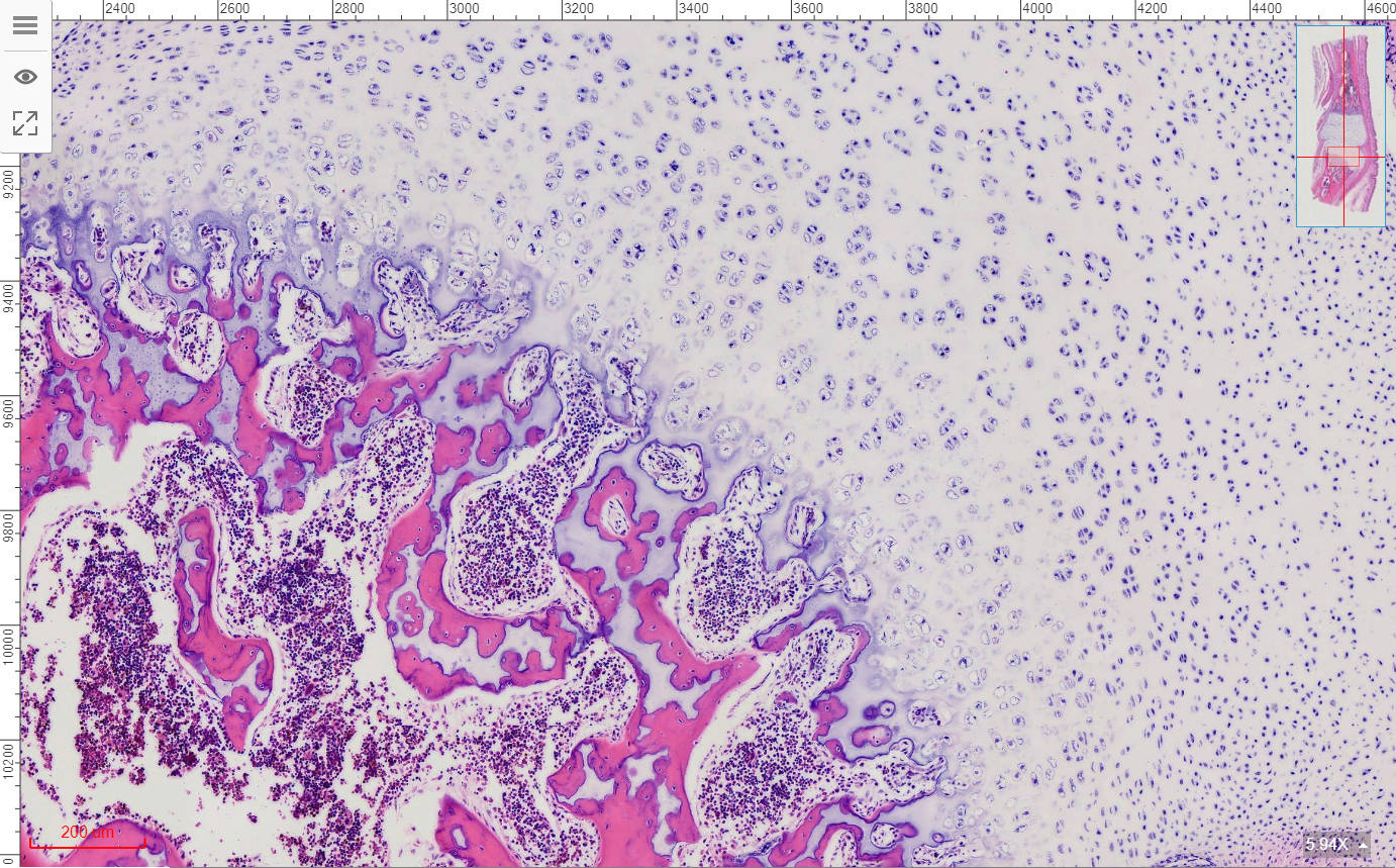
Zone of Hypertrophy – chondrocytes cease dividing, grow in size, and secrete cartilage matrix
Zone of Calcification – calcification of the cartilage matrix inhibits diffusion of nutrients and the chondrocytes undergo apoptosis
Zone of Ossification – osteoprogenitor cells migrate into the cavities with new blood vessels, differentiate into osteoblasts, and deposit new bone (eosinophilic) on the scaffold of calcified cartilage (basophilic)
From Histologyguide.
Scientific name: Endochondral Bone Development
Category: histology slides
Description of the Endochondral Bone Development:
- Size: 76.2*25.4mm
- Thickness: 7 um, sagittal section
- Stain: H.E. stain
- Show: different zone of Endochondral Bone Development
- Storage: long-lasting
- MOQ: 1PC
Other fixed kits:
- 100pcs/set, https://www.ihappysci.com/product/100pcs-human-histology-slides/
- 80pcs/set, https://www.ihappysci.com/product/80pcs-animal-histology-slides/
About Payment
Online orders only accept PayPal (Credit card available).
Contact us if you need to pay via Bank transfer, MoneyGram, Western Union, WeChat Pay, or Alipay.

About Shipment
We can ship worldwide by popular express.
Normally your order will be processed and shipped within 3 workdays.
We will contact you and confirm the estimated shipping date if it needs more time.

About Customization work
If the items you need are not on the list, please send us your list and get the quotation within 12 hours.
As a prepared microscope slides manufacturer, we also provide OEM service to customize your specially prepared slide sets.
Practical knowledge of Prepared microscope slides
C.S. = Cross Section: slide shows a thin section through the transverse plane of an organism or tissue.
L.S. = Longitudinal Section: slide shows a vertical section of the organism along the longest plane.
C.S. & L.S. = Cross Section and Longitudinal Section on the same slide for comparison
W.M. = Whole Mount: slide shows an entire organism or structure, as indicated, is preserved on the slide
About us:
Microscope Prepared Slides Manufacturer from China
What we have is shown following:
| Different Categories | Quantity |
| Botany | 868 kinds |
| Histology | 375 kinds |
| Zoology | 459 kinds |
| Human Pathology | 300 kinds |
| Mitosis&Meiosis | 66 kinds |
| Embryology | 33 kinds |
| Microbiology | 100 kinds |
| Cell Biology&Genetics | 64 kinds |
| Botany Pathology | 373 kinds |
| Oral Pathology | 95 kinds |
| Hematology | 59 kinds |
| Animal Pathology | 68 kinds |
| Petrology, rocks thin sections | 191 kinds |
Maybe the items you need are not on our website, please send us your list and get the quotation within 12 hours.
As a prepared microscope slides manufacturer, we provide OEM service.
For more order and customs information please visit the FAQ.
Write us an Email: [email protected];
Message us via WhatsApp: +86 18838161683.


Reviews
There are no reviews yet.